Ankle Posterior Drawer Test
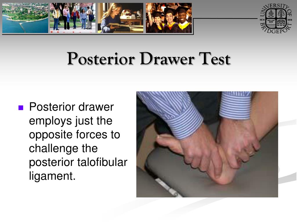
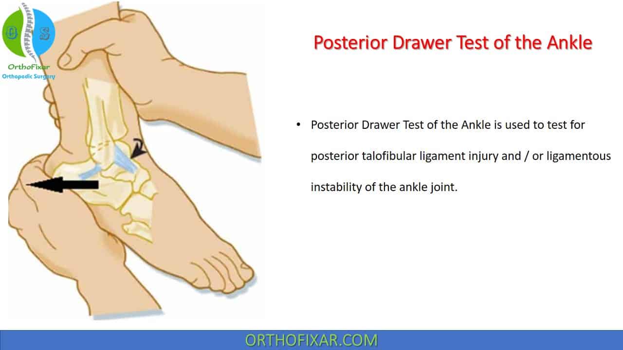
Ankle Posterior Drawer Test - In particular, it prevents the talus bone from moving too far forward. Joint laxity indicates a positive test. The examiner attempts to translate the fibula from anterior to posterior. Web the anterior drawer test can be used to assess the integrity of the anterior talofibular ligament 8 , and the inversion stress test can be used to assess the integrity of the calcaneofibular. With the ankle joint held at 10 to 15° of plantar flexion, the examiner grasps around the heel with one hand and stabilizes the tibia from the anterior side with the other. Frost and hanson 7 described the posterior drawer test using the same patient and clinician positioning as that used for the anterior drawer test. Web special test:posterior drawer test (ankle) procedure: This test assesses for a tear of the posterior cruciate ligament (pcl). Web testing for:posterior talofibular ligament injury and/or ligamentous instability procedure: Click here to check it out:. With the patient supine, flex the patient’s knee to 90º and place their foot flat on the table. In the normal ankle, there is a firm end point and little movement. Web the painful conditions of the ankle and foot are very common presentations and most commonly caused by trauma or injury related to sport activities. We have a new website!! Web about press copyright contact us creators advertise developers terms privacy policy & safety how youtube works test new features nfl sunday ticket press copyright. At the attachments of the medial and lateral ligaments; Web ankle posterior drawer test is performed with the patient lies supine with the knee slightly flexed to neutralize the pull of the gastrocnemius muscle. Click here to check it out:. This test assesses for a tear of the posterior cruciate ligament (pcl). Click here to jump onto our email list. In acute injuries, the eversion stress test may be of limited clinical value. At the attachments of the medial and lateral ligaments; Web the posterior drawer test is used to assess the integrity of the posterior cruciate ligament. Presence of sulcus, pain, or excessive posterior translation of the talus, indicating ligamentous laxity or rupture negative: Click here to jump onto. Web about press copyright contact us creators advertise developers terms privacy policy & safety how youtube works test new features nfl sunday ticket press copyright. Web anterior drawer test: On the medial, lateral, posterior and anterior part of the lower leg and the around calcaneus; The anterior drawer test helps evaluate ankle injuries, particularly from outward rolls that may stretch. With the knee flexed to 90 degrees and the foot stabilized (often the examiner sits on the patient's foot), the proximal tibia is grasped firmly with both hands and the tibia is forcibly pushed posteriorly, noting any laxity compared with the other side. Anterior drawer of the ankle. Click here to check it out:. The patient is positioned to promote. This test helps to rule in a positive posterior talofibular ligament sprain. With the ankle joint held at 10 to 15° of plantar flexion, the examiner grasps around the heel with one hand and stabilizes the tibia from the anterior side with the other. •patient is supine with foot relaxed •examiner stabilizes tibia and fibula with one hand •with the. In the normal ankle, there is a firm end point and little movement. You’ll lie on your back and a provider will move your lower leg to check how far your knee moves. Stabilize the ankle with your hip and push the proximal tibia posteriorly (away from you). With the ankle joint held at 10 to 15° of plantar flexion,. Web the anterior drawer test checks the health of the anterior talofibular ligament (atfl), a key ligament that helps keep the ankle joint stable. The anterior drawer test for ankle. Normal end feel and limited posterior translation, indicating intact ligaments. Patient is supine with foot relaxedtherapist stabilizes tibia and fibula with one handwith the patient’s foot plantar flexed to 20. Anterior drawer of the ankle. Under greatest strain in ankle dorsiflexion and acts to limit posterior talar displacementwithin the mortise as well as talar external rotation. Web anterior drawer test: Web the posterior drawer test is used to assess the integrity of the posterior cruciate ligament. With the ankle joint held at 10 to 15° of plantar flexion, the examiner. This test helps to rule in a positive posterior talofibular ligament sprain. In the normal ankle, there is a firm end point and little movement. A sensitivity of 52% has been reported in a single study for the inversion talar tilt test. The anterior drawer test helps evaluate ankle injuries, particularly from outward rolls that may stretch or tear the. Web about press copyright contact us creators advertise developers terms privacy policy & safety how youtube works test new features nfl sunday ticket press copyright. In the normal ankle, there is a firm end point and little movement. Web the anterior drawer test checks the health of the anterior talofibular ligament (atfl), a key ligament that helps keep the ankle. •patient is supine with foot relaxed •examiner stabilizes tibia and fibula with one hand •with the patient’s foot plantar flexed to 20 degrees, the examiner holds the patient’s calcaneus with other hand then distracts the calcaneus from the tibia and fibula ( by slowly pulling the calcanues inferiorly) Web ankle posterior drawer test is performed with the patient lies supine. Test for “high” (syndesmotic) ankle sprain (see below) imaging. Web the painful conditions of the ankle and foot are very common presentations and most commonly caused by trauma or injury related to sport activities. Web about press copyright contact us creators advertise developers terms privacy policy & safety how youtube works test new features nfl sunday ticket press copyright. Stabilize the ankle with your hip and push the proximal tibia posteriorly (away from you). With the knee flexed to 90 degrees and the foot stabilized (often the examiner sits on the patient's foot), the proximal tibia is grasped firmly with both hands and the tibia is forcibly pushed posteriorly, noting any laxity compared with the other side. The anterior drawer test for ankle. Joint laxity indicates a positive test. Patient is supine with foot relaxedtherapist stabilizes tibia and fibula with one handwith the patient’s foot plantar flexed to 20 degrees, the therapist holds the patient’s calcaneus with other hand then distracts the calcaneus from the tibia and fibula (by slowly pulling the. In acute injuries, the eversion stress test may be of limited clinical value. Anterior drawer of the ankle. Want to join the oep community? Healthcare providers sometimes call this a posterior drawer test, and some perform it at the same time as an anterior drawer test. A sensitivity of 52% has been reported in a single study for the inversion talar tilt test. With the patient supine, flex the patient’s knee to 90º and place their foot flat on the table. With the ankle joint held at 10 to 15° of plantar flexion, the examiner grasps around the heel with one hand and stabilizes the tibia from the anterior side with the other. Web 5.apply posterior pressure on the calcaneus and talus, and overpressure at the end of the passive range.
Posterior Drawer Test, PCL Injury Tests —

Posterior Drawer Test Posterior Cruciate Ligament YouTube
PPT Ankle and Foot Orthopaedic Tests Orthopedics and Neurology DX 612

Posterior drawer test for the ankle YouTube

Posterior Drawer test of ankle YouTube

Ankle Posterior Drawer Test YouTube

Posterior Drawer Test I 후거비인대(PTFL) 염좌 평가 I ankle10 YouTube
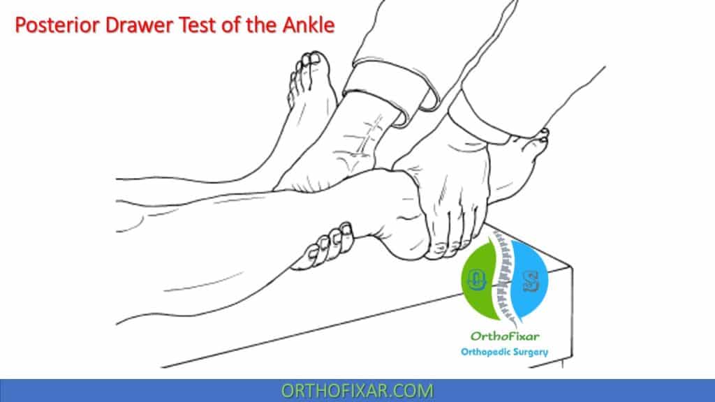
Posterior Drawer Test Of The Ankle 2024
Posterior Drawer Test Ankle
Posterior Drawer Test Of The Ankle 2024
This Test Helps To Rule In A Positive Posterior Talofibular Ligament Sprain.
Web Special Test:posterior Drawer Test (Ankle) Procedure:
At The Attachments Of The Medial And Lateral Ligaments;
Web This Video Demonstrates How To Perform A Posterior Drawer Test For The Ankle.
Related Post: