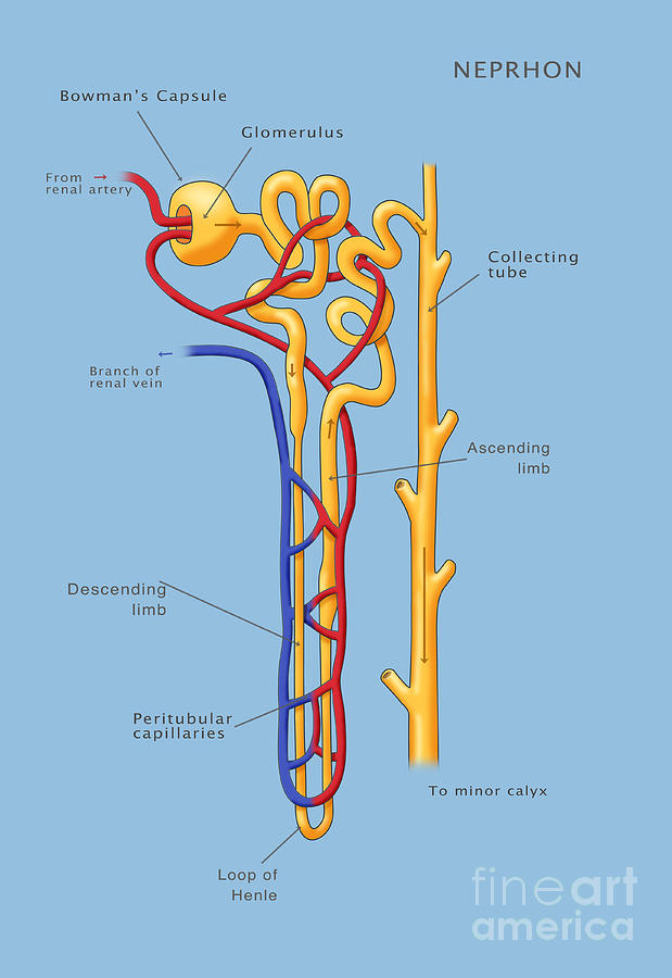
Drawing Of A Nephron
Drawing Of A Nephron - Ultrafiltration occurs when blood pressure forces water and other. Web learn for free about math, art, computer programming, economics, physics, chemistry, biology, medicine, finance, history, and more. Web this is the head of the nephron and it is where urine first forms. A nephron consists of two main parts: It look somewhat like a roll of yarn. Renal corpuscles are located in the renal cortex, while their tubular systems extend into the medulla. Web drawing of a nephron. Labels on the kidney cross section show where unfiltered blood enters, filtered blood leaves, and urine exits. You can learn how to draw and label the parts of nephron diagram, just watching this vide. It produces concentrated urine by creating an ultrafiltrate from blood. A nephron is the basic unit of structure in the kidney. Renal corpuscles are located in the renal cortex, while their tubular systems extend into the medulla. On the nephron, the glomerulus, tubule, and collecting duct are labeled along with where unfiltered blood enters, filtered blood exits, and urine exits. Web drawing of a nephron. The nephron functions through ultrafiltration. The renal corpuscle has two components: Each nephron has basically two parts. It produces concentrated urine by creating an ultrafiltrate from blood. A renal corpuscle and its associated renal tubule system. Labels on the kidney cross section show where unfiltered blood enters, filtered blood leaves, and urine exits. Web this video tutorial demonstrates how to draw nephron step by step. The renal corpuscle is the filtering component and renal tubules carry the filtered liquid away. Web drawing of a nephron. Web image of a close up nephron and its place in the kidney. A nephron is the basic unit of structure in the kidney. The glomerulus is like a network of capillaries. Web learn for free about math, art, computer programming, economics, physics, chemistry, biology, medicine, finance, history, and more. On the nephron, the glomerulus, tubule, and collecting duct are labeled along with where unfiltered blood enters, filtered blood exits, and urine exits. The most primitive nephrons are found in the kidneys of primitive. They are used by the kidney to separate substances from bodily fluids like ions, small molecules, and water, filtering out toxins and wastes from the blood and returning substances of interest back to the blood. Diagrams in cbse (ncert) class 10th biology explained in detailed and step wise method. The renal corpuscle is the filtering component and renal tubules carry. Web each nephron contains arenal corpuscle, which is the initial component that filters the blood, and a renal tubule that processes and carries the filtered fluid to the system of calices. This video helps you to draw science diagrams with great ease and clarity.nephron is t. Diagrams in cbse (ncert) class 10th biology explained in detailed and step wise method.. Renal corpuscles are located in the renal cortex, while their tubular systems extend into the medulla. The capsule and tubule are connected and are. The first and primary sort of water and ion reabsorption in the kidney, where all glucose in the blood is reabsorbed.; Glomerulus, bowman’s capsule, proximal and distal tubules, loop of henle, collecting duct and capillaries. A. Web learn for free about math, art, computer programming, economics, physics, chemistry, biology, medicine, finance, history, and more. Web drawing of a nephron. The renal corpuscle has two components: Nephrons are complexes of a few components: Renal corpuscles are located in the renal cortex, while their tubular systems extend into the medulla. Web how to draw nephron easily step by step for class 10 students by using some easy tricks which helps to draw the structure of nephron in examination of class. The first and primary sort of water and ion reabsorption in the kidney, where all glucose in the blood is reabsorbed.; A renal corpuscle and its associated renal tubule system.. A renal corpuscle and its associated renal tubule system. The nephron functions through ultrafiltration. Renal corpuscles are located in the renal cortex, while their tubular systems extend into the medulla. A small, intertwined group of capillaries within the nephrons of. The capsule and tubule are connected and are. Labels on the kidney cross section show where unfiltered blood enters, filtered blood leaves, and urine exits. A filtration unit, the renal corpuscle (composed of a glomerulus and bowman's capsule/glomerular capsule) that filters the blood, kidney tubules that allow adjustment. The bowman’s capsule, around the glomerulus, shapes like a funnel. Web image of a close up nephron and its place. This was designed to go with a larger unit on how the urinary system and kidneys help the body maintain water balance and homeostasis. Diagrams in cbse (ncert) class 10th biology explained in detailed and step wise method. Web image of a close up nephron and its place in the kidney. You can learn how to draw and label the. Nephrons are functional units located in the kidneys responsible for forming urine. A nephron is used separate to water, ions and small molecules from the blood, filter out wastes and toxins, and return needed molecules to the blood. Web ‘nephron diagram || how to draw and label the parts of a nephron’ is demonstrated in this video tutorial step by step. Web this is the head of the nephron and it is where urine first forms. Nephrons are complexes of a few components: Comprises the rest 15% of the nephrons in the human kidney, they start low in the cortex near the medulla and. The video provides a detailed overview of the kidney's smallest functional unit, the nephron. Most instructors don't draw in all the details of the capillary network because it covers up the nephron. The first and primary sort of water and ion reabsorption in the kidney, where all glucose in the blood is reabsorbed.; It produces concentrated urine by creating an ultrafiltrate from blood. Web this video tutorial demonstrates how to draw nephron step by step. Web drawing of a nephron. This was designed to go with a larger unit on how the urinary system and kidneys help the body maintain water balance and homeostasis. Glomerulus, bowman’s capsule, proximal and distal tubules, loop of henle, collecting duct and capillaries. Web nephron the nephron is the functional unit of the kidney. Web students can also color the image to identify the major structures of the nephron: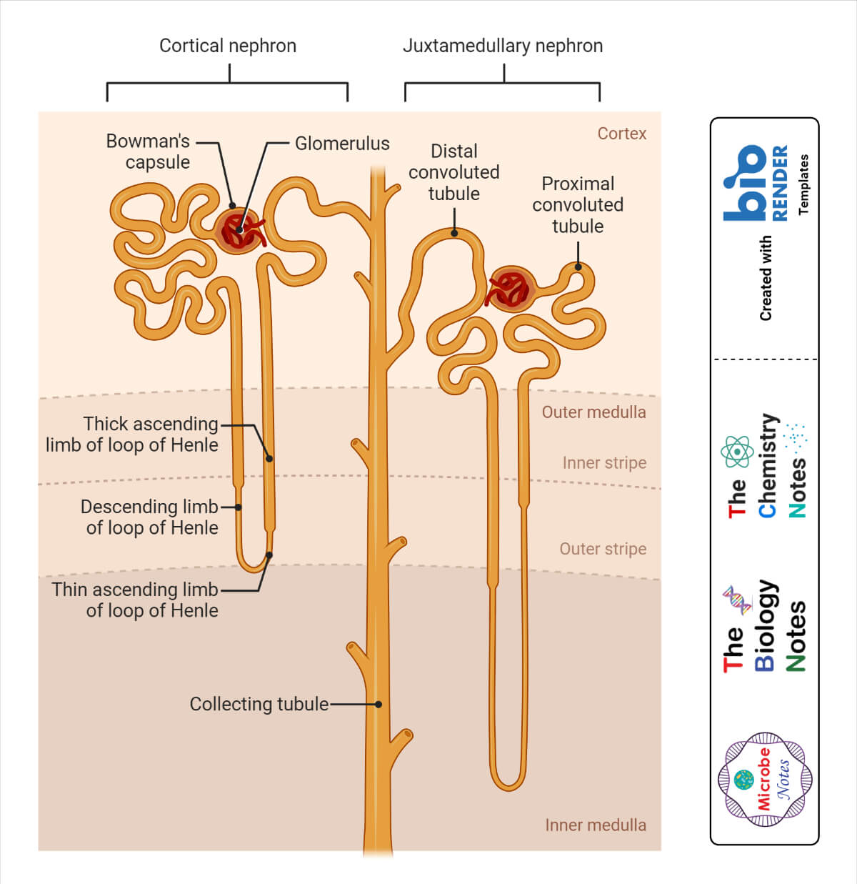
Nephron Definition, Structure, Physiology, Functions
Nephron Definition, Parts, Structure, & Functions, with Diagram

How to draw easy diagram of nephron step by step for beginners YouTube

How To Draw Nephron step by step for beginners YouTube
Nephron The Functioning Unit of The Kidney Interactive Biology, with
Well labelled diagram of nephron.

Anatomy And Physiology Of The Nephron My Endo Consult

How To Draw Structure Of Nephron Diagram Step By Step For Beginners
Nephron Of The Kidney, Illustration 1 Photograph by Monica Schroeder

How to draw Nephron diagram easily step by step for beginners class 10
Cortical Nephrons Start High In The Cortex That Does Not Penetrate Deep Into The Medulla And Has A Characteristic Short Henle’s Loop.
Each Nephron Has Basically Two Parts.
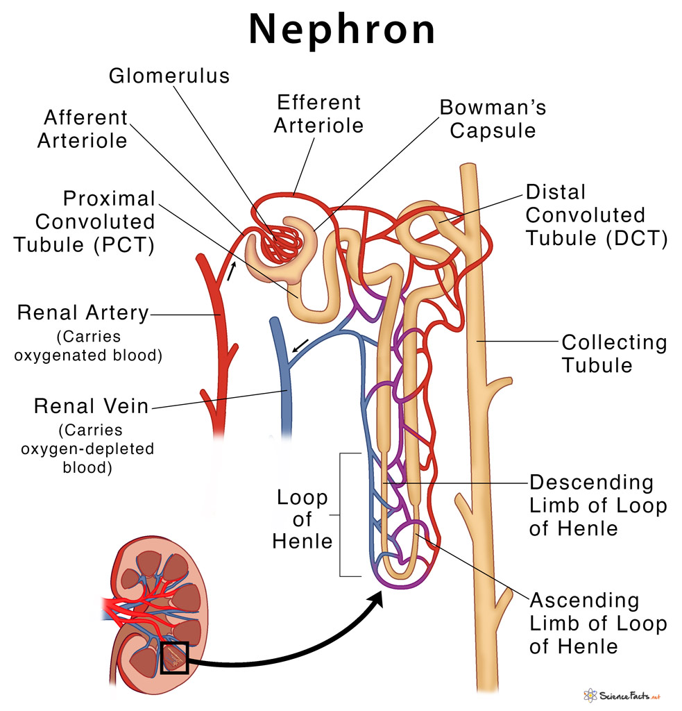
The Renal Corpuscle Is The Filtering Component And Renal Tubules Carry The Filtered Liquid Away.
The Glomerular (Bowman’s) Capsule In Which Sits The Glomerulus.
Related Post: